Copyright
©The Author(s) 2015.
World J Exp Med. May 20, 2015; 5(2): 140-153
Published online May 20, 2015. doi: 10.5493/wjem.v5.i2.140
Published online May 20, 2015. doi: 10.5493/wjem.v5.i2.140
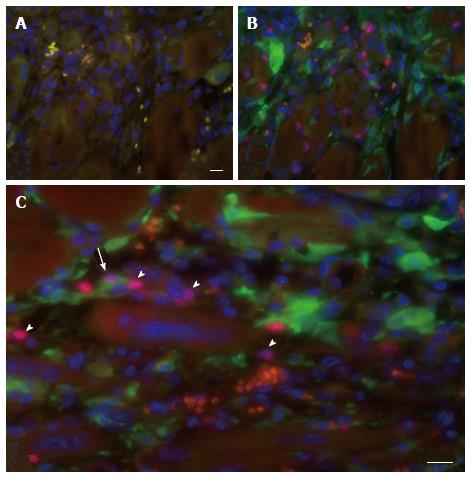
Figure 6 Contribution of host cells to myoregeneration in the muscle implant.
Immunofluorescence analysis of a fresh mouse implant excised 7 d after implantation in eGFP transgenic recipients. Host cell contribution was evaluated in tissue sections stained with the karyophilic fluorochrome Hoechst 33342 and antibodies specific for eGFP and Pax7. A: Negative control section incubated with Hoechst 33342 (blue), the secondary antibodies and Cy3-conjugated streptavidin; B: Section stained for Pax7 (red), eGFP (green) and Hoechst 33342 (blue); C: Example of a cell positive for both Pax7 and eGFP (arrow) situated at the periphery of the implant. Also present are cells only positive for Pax7 (arrowheads) or for GFP, blood vessels with erythrocytes (orange) and multinucleated myofibers (center of image). Scale bar for a and b is 50 μm. Panel (C) shows an electronic enlargement of an image recorded at the same magnification as (A) and (B).
- Citation: Garza-Rodea ASL, Boersma H, Dambrot C, Vries AA, Bekkum DWV, Knaän-Shanzer S. Barriers in contribution of human mesenchymal stem cells to murine muscle regeneration. World J Exp Med 2015; 5(2): 140-153
- URL: https://www.wjgnet.com/2220-315X/full/v5/i2/140.htm
- DOI: https://dx.doi.org/10.5493/wjem.v5.i2.140


