Copyright
©2013 Baishideng.
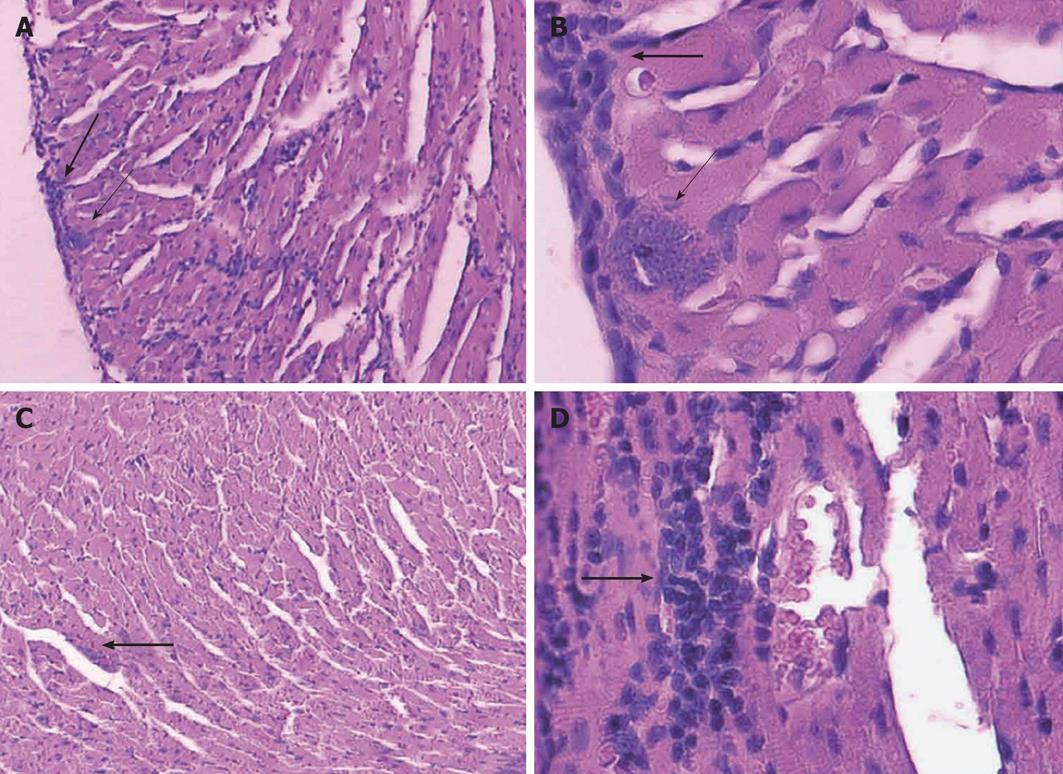
Figure 2 Histological studies: all sections were stained with hematoxylin and eosin.
Histological sections of heart. A, B: Control groups show nests of amastigotes (thin arrows) and mononuclear cell infiltration (thick arrow); C, D: Representative sections from mice vaccinated with Trypanosoma cruzi. Infected mice show focal mononuclear cell infiltrates (thick arrows). No amastigote nest was observed (A, C: 100 ×; B, D: 400 ×).
- Citation: Basso B. Modulation of immune response in experimental Chagas disease. World J Exp Med 2013; 3(1): 1-10
- URL: https://www.wjgnet.com/2220-315X/full/v3/i1/1.htm
- DOI: https://dx.doi.org/10.5493/wjem.v3.i1.1


