Copyright
©2012 Baishideng.
World J Exp Med. Aug 20, 2012; 2(4): 78-85
Published online Aug 20, 2012. doi: 10.5493/wjem.v2.i4.78
Published online Aug 20, 2012. doi: 10.5493/wjem.v2.i4.78
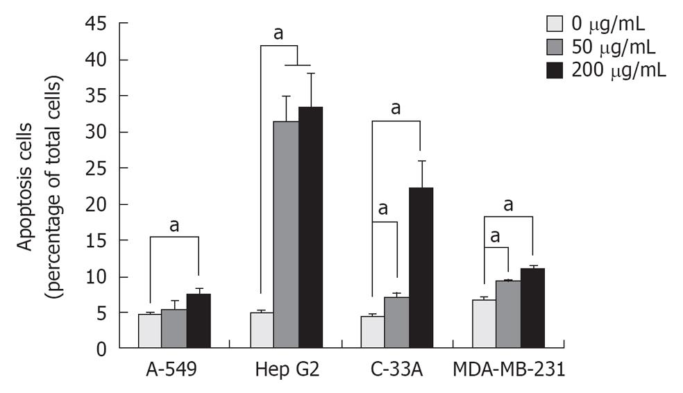
Figure 4 Detection of longan seed extract-induced apoptotic cells.
Phosphatidylserine is usually distributed in the inner fleet of the plasma membrane. When cells are undergoing apoptosis, this phospholipid translocates to the outer fleet, and can be recognized by a bacteria glycoprotein named annexin V. We used annexin V conjugated with fluorescein isothiocyanate (FITC) as an apoptosis indicator and analyzed the apoptotic cells by flow cytometry. Briefly, 50 and 200 μg/mL longan seed extract-treated cells were incubated at 37 °C for 48 h. The treated cells were then suspended and stained with annexin V conjugated with FITC. Ten thousand cells were analyzed by flow cytometry using FL-1 as the parameter. Data are taken from the averages of three independent experiments and expressed as the mean ± SD. aP < 0.05.
- Citation: Lin CC, Chung YC, Hsu CP. Potential roles of longan flower and seed extracts for anti-cancer. World J Exp Med 2012; 2(4): 78-85
- URL: https://www.wjgnet.com/2220-315X/full/v2/i4/78.htm
- DOI: https://dx.doi.org/10.5493/wjem.v2.i4.78


