Copyright
©2012 Baishideng.
World J Exp Med. Aug 20, 2012; 2(4): 78-85
Published online Aug 20, 2012. doi: 10.5493/wjem.v2.i4.78
Published online Aug 20, 2012. doi: 10.5493/wjem.v2.i4.78
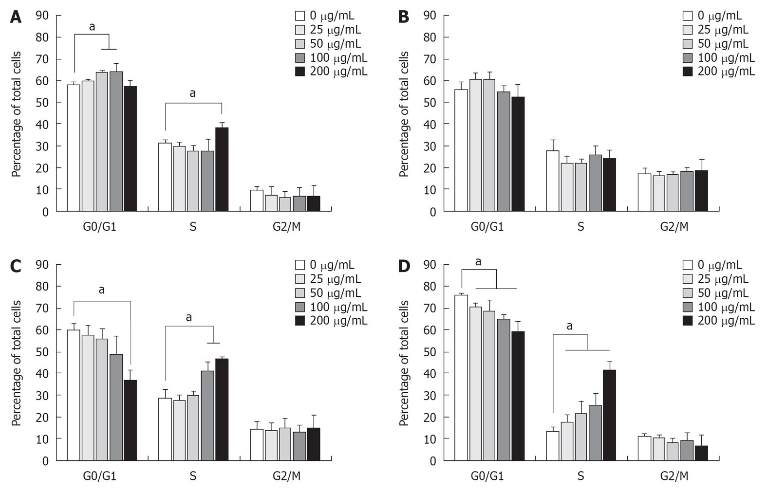
Figure 2 Cell cycle analysis of longan seed extract -treated cancer cells.
About 1 × 10⁶ cells in 10 mL medium in 100 mm plate were treated with increasing concentrations of longan seed extract as indicated, then incubated at 37 °C for 48 h. The (A) A-549, (B) Hep G2, (C) C-33A and (D) MDA-MB-231 cells harvested by trypsinization were fixed in 70% alcohol at -20 °C for 2 h and then reconstituted in phosphate-buffered saline. The cells were stained with propidium iodide solution in the dark at room temperature for 30 min. The stained cells were then analyzed using the FL-2A parameter of a flow cytometer to obtain the DNA content of the cells, and the distribution in each cell-cycle phase was determined using Modfit software. Data are expressed as a percentage of the total cells. Data represent the average of three independent experiments, and are expressed as the mean ± SD. aP < 0.05.
- Citation: Lin CC, Chung YC, Hsu CP. Potential roles of longan flower and seed extracts for anti-cancer. World J Exp Med 2012; 2(4): 78-85
- URL: https://www.wjgnet.com/2220-315X/full/v2/i4/78.htm
- DOI: https://dx.doi.org/10.5493/wjem.v2.i4.78


