Copyright
©The Author(s) 2019.
World J Crit Care Med. Nov 19, 2019; 8(7): 106-119
Published online Nov 19, 2019. doi: 10.5492/wjccm.v8.i7.106
Published online Nov 19, 2019. doi: 10.5492/wjccm.v8.i7.106
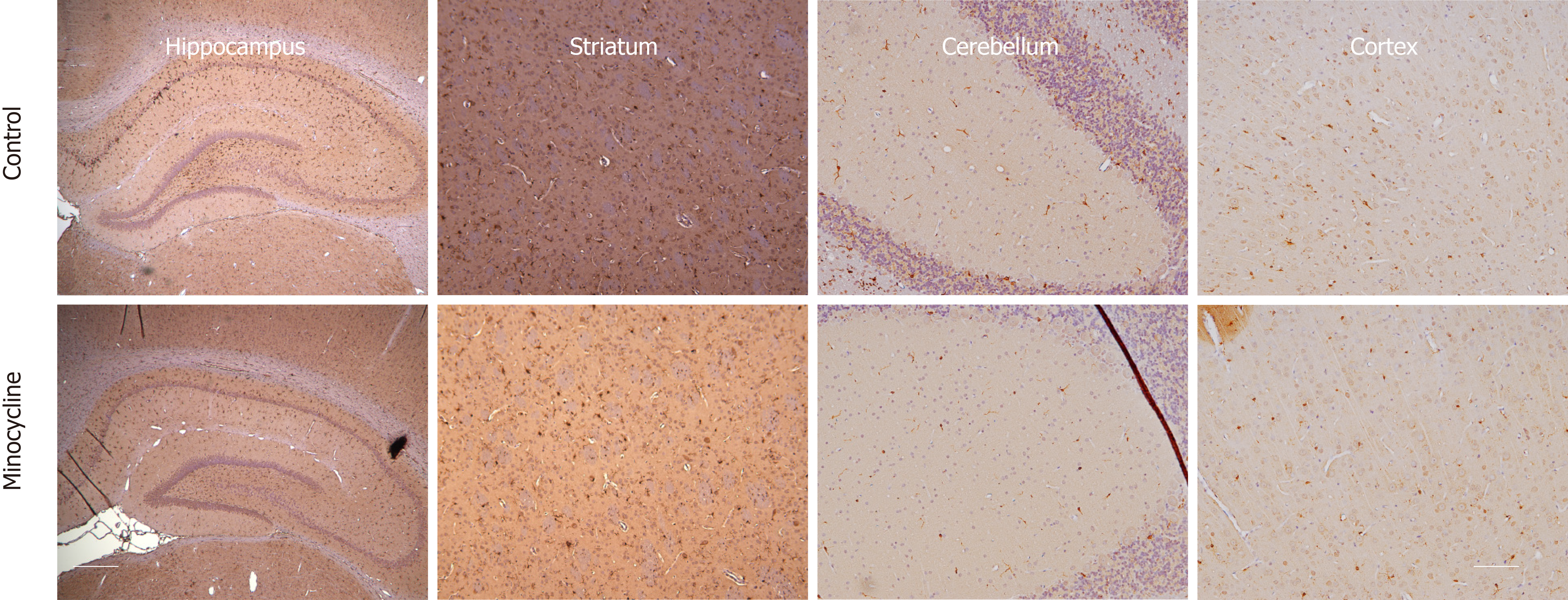
Figure 7 Representative samples of microglial activation and proliferation after 6 min ventricular fibrillation cardiac arrest at 72 h.
Sections are stained with hematoxylin. Brown staining is anti-Iba-1 staining, visualizing microglia, counterstained with diaminobenzamide. Magnification × 10. The scale bars in the far left and far right lower panels represent 10 µm.
- Citation: Janata A, Magnet IA, Schreiber KL, Wilson CD, Stezoski JP, Janesko-Feldman K, Kochanek PM, Drabek T. Minocycline fails to improve neurologic and histologic outcome after ventricular fibrillation cardiac arrest in rats. World J Crit Care Med 2019; 8(7): 106-119
- URL: https://www.wjgnet.com/2220-3141/full/v8/i7/106.htm
- DOI: https://dx.doi.org/10.5492/wjccm.v8.i7.106


