Copyright
©The Author(s) 2016.
World J Crit Care Med. Feb 4, 2016; 5(1): 7-11
Published online Feb 4, 2016. doi: 10.5492/wjccm.v5.i1.7
Published online Feb 4, 2016. doi: 10.5492/wjccm.v5.i1.7
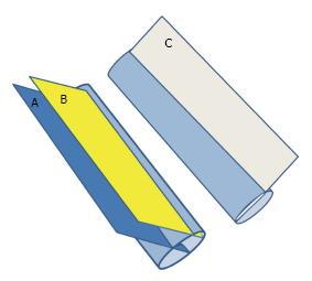
Figure 2 Three dimensional diagram showing the longitudinal ultrasound measurement of the antero-posterior diameter.
Measurements depend on the site and angle at which it crosses the IVC. Section A is the proper one as it crosses the IVC vertically at the midpoint. Section B crosses the IVC vertically but peripherally and gives a false low measurement of the IVC diameter. Section C crosses the IVC obliquely and gives a false high measurement of the IVC diameter. IVC: Inferior vena cava.
- Citation: Abu-Zidan FM. Optimizing the value of measuring inferior vena cava diameter in shocked patients. World J Crit Care Med 2016; 5(1): 7-11
- URL: https://www.wjgnet.com/2220-3141/full/v5/i1/7.htm
- DOI: https://dx.doi.org/10.5492/wjccm.v5.i1.7


