Copyright
©2014 Baishideng Publishing Group Inc.
World J Crit Care Med. Nov 4, 2014; 3(4): 80-94
Published online Nov 4, 2014. doi: 10.5492/wjccm.v3.i4.80
Published online Nov 4, 2014. doi: 10.5492/wjccm.v3.i4.80
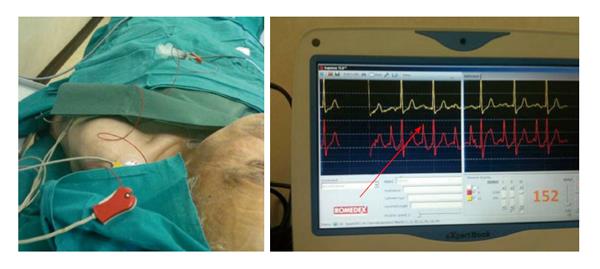
Figure 7 The electrocardiographic method (intracavitary electrocardiography).
The red arrow shows the maximal height of P-wave detectable when the catheter tip is at cavo-atrial junction (intracavitary EKG = red line, lead II). The yellow line is the surface EKG (lead III). EKG: Electrocardiography.
- Citation: Cotogni P, Pittiruti M. Focus on peripherally inserted central catheters in critically ill patients. World J Crit Care Med 2014; 3(4): 80-94
- URL: https://www.wjgnet.com/2220-3141/full/v3/i4/80.htm
- DOI: https://dx.doi.org/10.5492/wjccm.v3.i4.80


