Copyright
©The Author(s) 2024.
World J Crit Care Med. Jun 9, 2024; 13(2): 93206
Published online Jun 9, 2024. doi: 10.5492/wjccm.v13.i2.93206
Published online Jun 9, 2024. doi: 10.5492/wjccm.v13.i2.93206
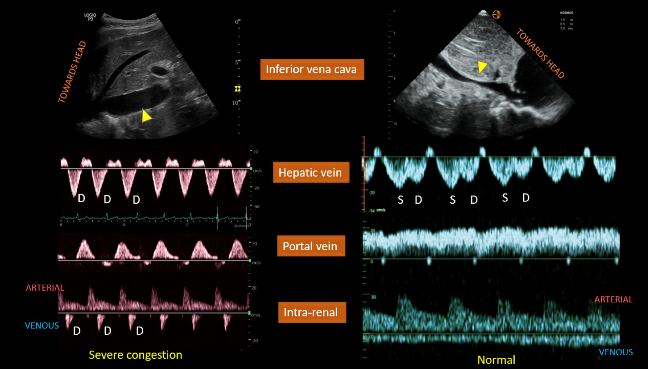
Figure 4 Left panel.
Plethoric inferior vena cava (IVC) (arrowhead), only diastolic (D) wave below-the-baseline on hepatic vein Doppler, pulsatile portal vein Doppler, and D-only or monophasic renal parenchymal vein Doppler obtained from a patient with fluid overload and severe venous congestion; right panel: normal-appearing IVC with < 2 cm diameter (arrowhead) and normal venous waveforms. Citation: Koratala A, Ronco C, Kazory A. Diagnosis of Fluid Overload: From Conventional to Contemporary Concepts. Cardiorenal Med. 2022; 12: 141-154. Copyright ©Silverchair Publisher 2022. Published by Karger Publishers.
- Citation: Khan AA, Saeed H, Haque IU, Iqbal A, Du D, Koratala A. Point-of-care ultrasonography spotlight: Could venous excess ultrasound serve as a shared language for internists and intensivists? World J Crit Care Med 2024; 13(2): 93206
- URL: https://www.wjgnet.com/2220-3141/full/v13/i2/93206.htm
- DOI: https://dx.doi.org/10.5492/wjccm.v13.i2.93206


