Copyright
©The Author(s) 2024.
World J Crit Care Med. Jun 9, 2024; 13(2): 91212
Published online Jun 9, 2024. doi: 10.5492/wjccm.v13.i2.91212
Published online Jun 9, 2024. doi: 10.5492/wjccm.v13.i2.91212
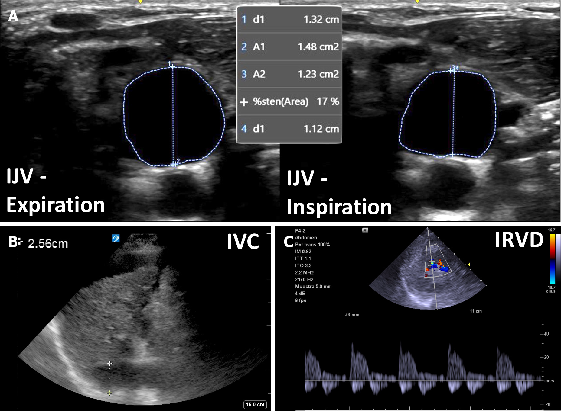
Figure 3 An example of venous congestion in cirrhosis.
A: Plethoric internal jugular vein with less than 25% antero-posterior inspiratory collapse; B: Plethoric inferior vena cava (> 2.0 cm); C: Biphasic intra-renal venous Doppler. IJV: Internal jugular vein; IVC: Inferior vena cava; IRVD: Intra-renal venous Doppler.
- Citation: Aguirre-Villarreal D, Leal-Villarreal MAJ, García-Juárez I, Argaiz ER, Koratala A. Sound waves and solutions: Point-of-care ultrasonography for acute kidney injury in cirrhosis. World J Crit Care Med 2024; 13(2): 91212
- URL: https://www.wjgnet.com/2220-3141/full/v13/i2/91212.htm
- DOI: https://dx.doi.org/10.5492/wjccm.v13.i2.91212


