Copyright
©The Author(s) 2022.
World J Crit Care Med. Sep 9, 2022; 11(5): 335-341
Published online Sep 9, 2022. doi: 10.5492/wjccm.v11.i5.335
Published online Sep 9, 2022. doi: 10.5492/wjccm.v11.i5.335
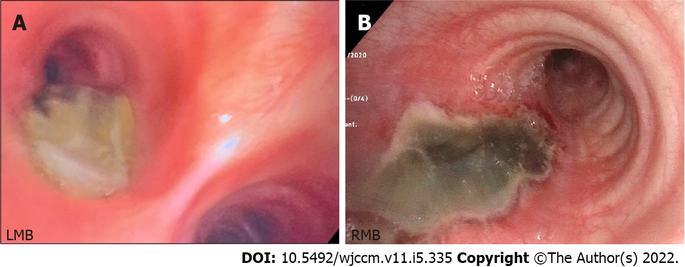
Figure 2 Flexible bronchoscopy.
A: Day 1: Two-centimeter bronchoesophageal fistula (asterisk) with adjacent yellow tinged devitalized mucosa on the posterior wall of left main bronchi; B: Day 8: Further delineation of fistulous track (asterisk) with necrotic mucosa and well-defined borders. LMB: Left main bronchi; RMB: Right main bronchi.
- Citation: Lagrotta G, Ayad M, Butt I, Danckers M. Cardiac arrest due to massive aspiration from a broncho-esophageal fistula: A case report. World J Crit Care Med 2022; 11(5): 335-341
- URL: https://www.wjgnet.com/2220-3141/full/v11/i5/335.htm
- DOI: https://dx.doi.org/10.5492/wjccm.v11.i5.335


