Copyright
©The Author(s) 2021.
World J Crit Care Med. Nov 9, 2021; 10(6): 310-322
Published online Nov 9, 2021. doi: 10.5492/wjccm.v10.i6.310
Published online Nov 9, 2021. doi: 10.5492/wjccm.v10.i6.310
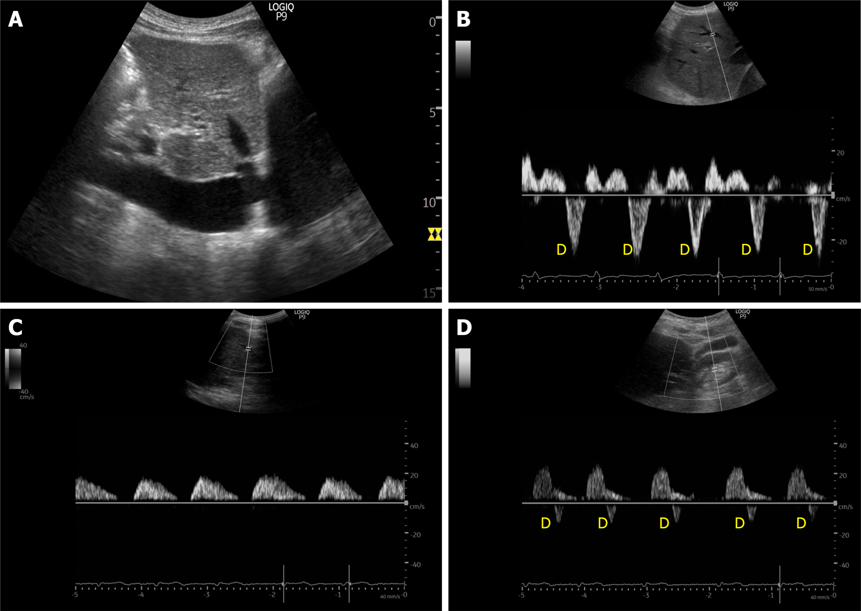
Figure 4 Example of ultrasound stigmata of severe venous congestion obtained from a patient with congestive heart failure exacerbation and tricuspid regurgitation.
A: Dilated inferior vena cava; B: Hepatic vein Doppler demonstrating only D-wave below the baseline; C: Pulsatile portal vein with flow pauses in between the cardiac cycles; D: Ontra-renal vein demonstrating only D-wave below the baseline.
- Citation: Galindo P, Gasca C, Argaiz ER, Koratala A. Point of care venous Doppler ultrasound: Exploring the missing piece of bedside hemodynamic assessment. World J Crit Care Med 2021; 10(6): 310-322
- URL: https://www.wjgnet.com/2220-3141/full/v10/i6/310.htm
- DOI: https://dx.doi.org/10.5492/wjccm.v10.i6.310


