Copyright
©The Author(s) 2021.
World J Crit Care Med. Sep 9, 2021; 10(5): 204-219
Published online Sep 9, 2021. doi: 10.5492/wjccm.v10.i5.204
Published online Sep 9, 2021. doi: 10.5492/wjccm.v10.i5.204
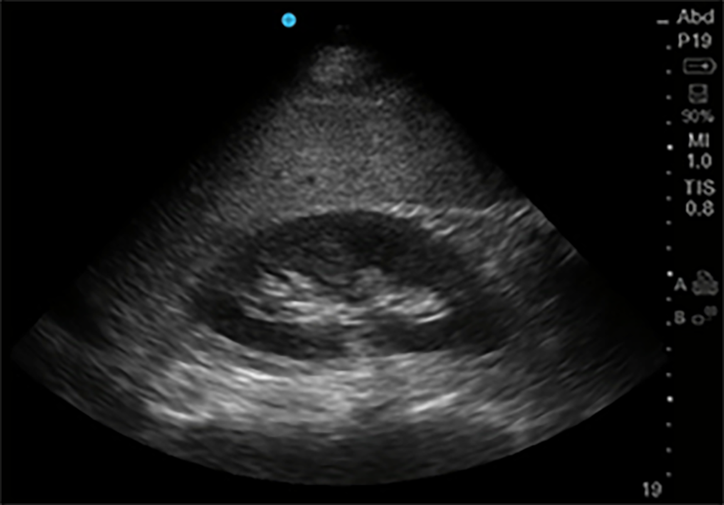
Figure 10 Kidney in its Longitudinal axis.
Phased array probe (1-5 MHz) in “Abdominal” preset placed with probe marker facing cephalad in right mid-axillary location. In this normal ultrasound, the liver serves as an acoustic window, under which can be seen the thin hyperechoic kidney capsule, the hypoechoic parenchymal cortex, and the central hyperechoic renal sinus.
- Citation: Deshwal H, Pradhan D, Mukherjee V. Point-of-care ultrasound in a pandemic: Practical guidance in COVID-19 units. World J Crit Care Med 2021; 10(5): 204-219
- URL: https://www.wjgnet.com/2220-3141/full/v10/i5/204.htm
- DOI: https://dx.doi.org/10.5492/wjccm.v10.i5.204


