Copyright
©The Author(s) 2021.
World J Crit Care Med. Sep 9, 2021; 10(5): 204-219
Published online Sep 9, 2021. doi: 10.5492/wjccm.v10.i5.204
Published online Sep 9, 2021. doi: 10.5492/wjccm.v10.i5.204
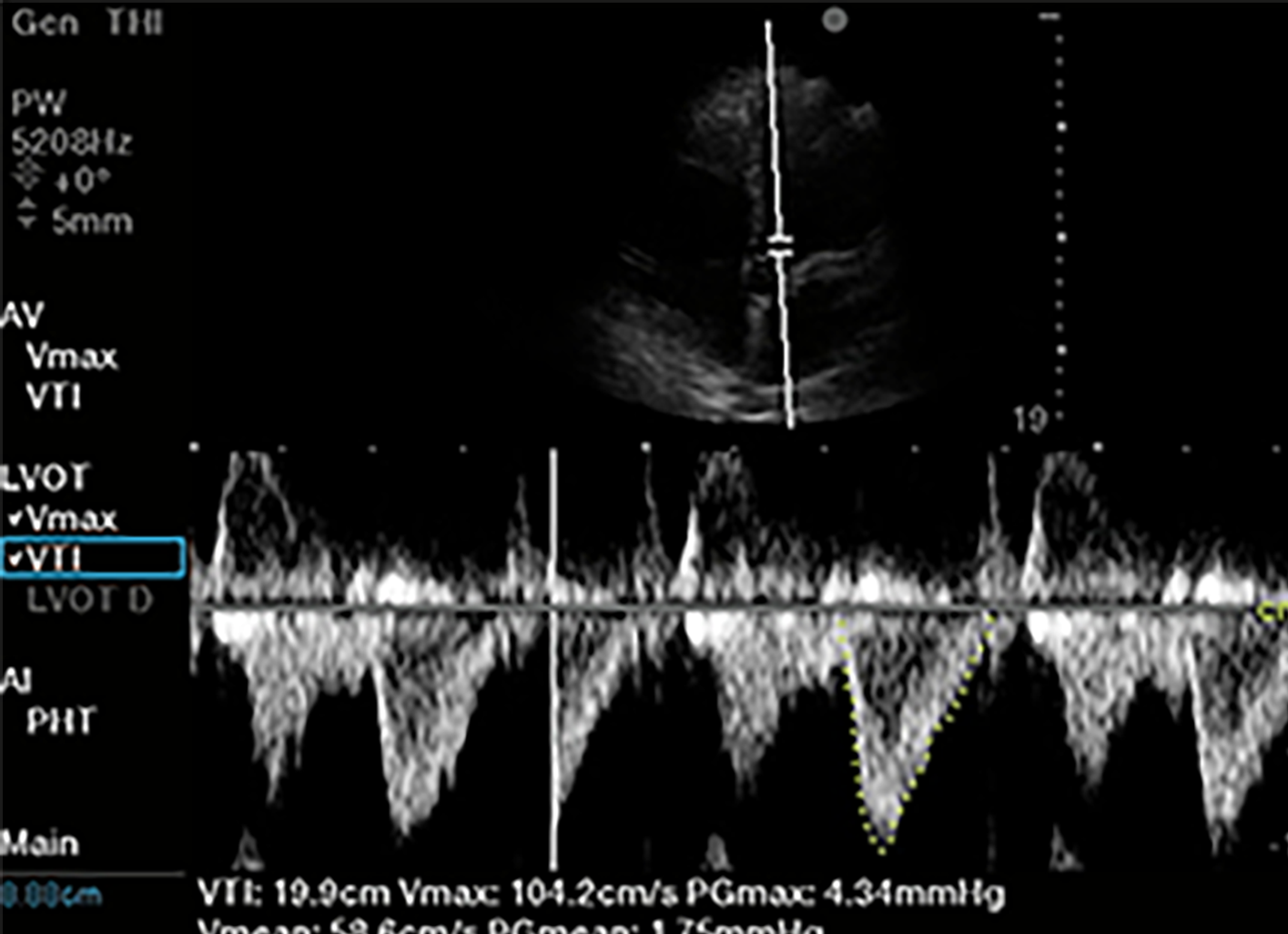
Figure 7 Left ventricular outflow tract velocity time integral.
Of 1-5 MHz phased array probe, apical 5 chamber view. Pulsed wave doppler selected, with sample volume placed 5 mm proximal to aortic valve in center of the left ventricular outflow tract. Notice narrow signal with rapid upstroke in velocities, with end-systolic click terminating flow signal. In this case traced velocity time integral was 19.9 cm.
- Citation: Deshwal H, Pradhan D, Mukherjee V. Point-of-care ultrasound in a pandemic: Practical guidance in COVID-19 units. World J Crit Care Med 2021; 10(5): 204-219
- URL: https://www.wjgnet.com/2220-3141/full/v10/i5/204.htm
- DOI: https://dx.doi.org/10.5492/wjccm.v10.i5.204


