Copyright
©2012 Baishideng.
World J Crit Care Med. Aug 4, 2012; 1(4): 102-105
Published online Aug 4, 2012. doi: 10.5492/wjccm.v1.i4.102
Published online Aug 4, 2012. doi: 10.5492/wjccm.v1.i4.102
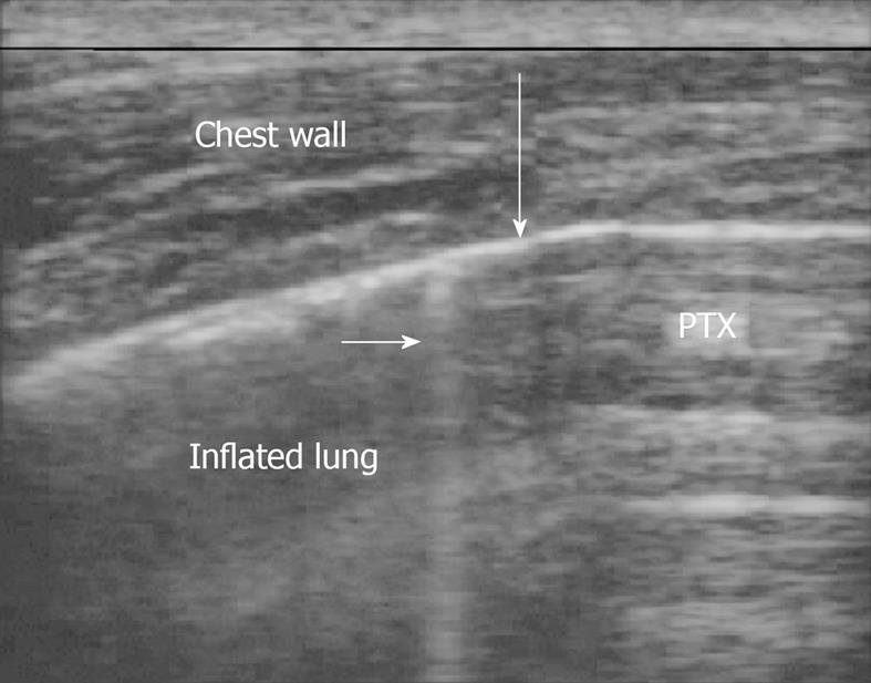
Figure 1 An ultrasound depiction of a pneumothorax is shown.
This is the lung point. To the right of the image air blocks visualization of typical lung artifacts. On the left, the visceral and parietal pleura are sliding past each other. The large arrow shows where they meet. The small arrow shows a B line, seen only in inflated portions of the lung.
- Citation: Blaivas M. Update on point of care ultrasound in the care of the critically ill patient. World J Crit Care Med 2012; 1(4): 102-105
- URL: https://www.wjgnet.com/2220-3141/full/v1/i4/102.htm
- DOI: https://dx.doi.org/10.5492/wjccm.v1.i4.102


