Copyright
©2012 Baishideng.
World J Crit Care Med. Jun 4, 2012; 1(3): 80-93
Published online Jun 4, 2012. doi: 10.5492/wjccm.v1.i3.80
Published online Jun 4, 2012. doi: 10.5492/wjccm.v1.i3.80
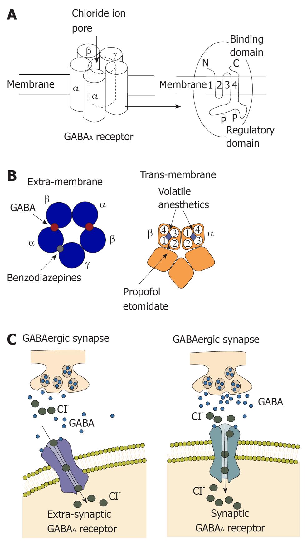
Figure 1 Structure and function of γ- aminobutyric acid type A receptor.
A: γ- aminobutyric acid type A (GABAA) receptors commonly contain two α subunits, two β subunits and one γ subunit. Chloride influx through the pore could hyperpolarize the postsynaptic membrane; B: Left: Extra-membrane region of GABAA receptor. The binding sites for GABA are located between α and β subunits and the binding site for benzodiazepines is located between γ and α subunits; Right: Trans-membrane region of GABAA receptor. Four trans-membrane segments form the α subunit. It has been shown that the trans-membrane segment of β subunit is the binding site for propofol and etomidate. This binding site is close to a binding site for volatile anesthetics; C: Activation of the GABAA receptor could increase conductance of the postsynaptic membrane and alter the potential of the membrane because of influx of chloridion. Synaptic receptors could detect GABA at mmol concentration to produce fast inhibitory postsynaptic potentials (IPSPs), and extra-synaptic receptors that detect GABA at μmol concentrations to produce slower IPSPs.
- Citation: Zhou C, Liu J, Chen XD. General anesthesia mediated by effects on ion channels. World J Crit Care Med 2012; 1(3): 80-93
- URL: https://www.wjgnet.com/2220-3141/full/v1/i3/80.htm
- DOI: https://dx.doi.org/10.5492/wjccm.v1.i3.80


