Copyright
©2014 Baishideng Publishing Group Co.
World J Surg Proced. Mar 28, 2014; 4(1): 13-20
Published online Mar 28, 2014. doi: 10.5412/wjsp.v4.i1.13
Published online Mar 28, 2014. doi: 10.5412/wjsp.v4.i1.13
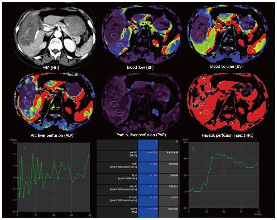
Figure 3 Transverse computed tomography perfusion functional maps of the blood volume, blood flow, portal-venous perfusion, arterial liver perfusion and hepatic perfusion index in a 49-year-old woman show a large alveolar echinococcosis lesion in the right lobe of the liver that has a distinct range of colors compared with the background liver parenchyma.
Perfusion values from an ROI drawn in the solid component without calcification of alveolar echinococcosis (ROI 1) and normal tissue (ROI 2) show lower blood flow, blood volume, arterial liver perfusion and portal-venous perfusion values compared with normal liver parenchyma.
- Citation: Kantarci M, Pirimoglu B, Kizrak Y. Diagnostic imaging and interventional procedures in a growing problem: Hepatic alveolar echinococcosis. World J Surg Proced 2014; 4(1): 13-20
- URL: https://www.wjgnet.com/2219-2832/full/v4/i1/13.htm
- DOI: https://dx.doi.org/10.5412/wjsp.v4.i1.13


