Copyright
©2014 Baishideng Publishing Group Co.
World J Surg Proced. Mar 28, 2014; 4(1): 13-20
Published online Mar 28, 2014. doi: 10.5412/wjsp.v4.i1.13
Published online Mar 28, 2014. doi: 10.5412/wjsp.v4.i1.13
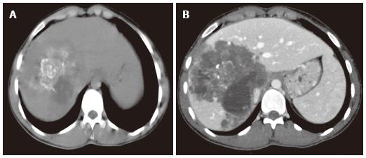
Figure 2 Alveolar echinococcosis in a 34-year-old man.
Axial unenhanced computed tomography (CT) image demonstrates an infiltrating tumor-like hepatic mass with irregular margins and heterogeneous contents, including scattered hyperattenuating foci of calcification and areas of hypoattenuation corresponding to necrosis and parasite tissue (A). Alveolar echinococcosis in a 29 year old man. Abdominal CT images obtained after the administration of intravenous contrast medium show a poor enhancement, hypoattenuating lesion in the portal venous phase (B).
- Citation: Kantarci M, Pirimoglu B, Kizrak Y. Diagnostic imaging and interventional procedures in a growing problem: Hepatic alveolar echinococcosis. World J Surg Proced 2014; 4(1): 13-20
- URL: https://www.wjgnet.com/2219-2832/full/v4/i1/13.htm
- DOI: https://dx.doi.org/10.5412/wjsp.v4.i1.13


