Copyright
©2014 Baishideng Publishing Group Co.
World J Surg Proced. Mar 28, 2014; 4(1): 13-20
Published online Mar 28, 2014. doi: 10.5412/wjsp.v4.i1.13
Published online Mar 28, 2014. doi: 10.5412/wjsp.v4.i1.13
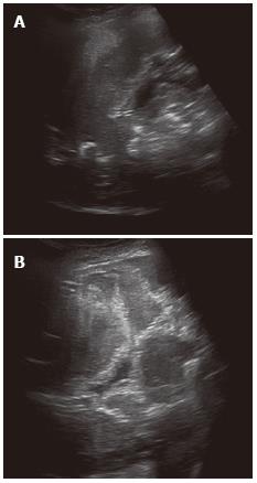
Figure 1 Alveolar echinococcosis in a 41-year-old woman.
Abdominal gray-scale ultrasonography (US) image shows a heterogeneous mass lesion in the right lobe of the liver. The mass is generally hypoechoic but contains hyperechoic foci of calcifications (A). Alveolar echinococcosis in a 38 year old woman. Abdominal gray-scale US image shows a heterogeneous, hyperechoic lesion without calcifications (B).
- Citation: Kantarci M, Pirimoglu B, Kizrak Y. Diagnostic imaging and interventional procedures in a growing problem: Hepatic alveolar echinococcosis. World J Surg Proced 2014; 4(1): 13-20
- URL: https://www.wjgnet.com/2219-2832/full/v4/i1/13.htm
- DOI: https://dx.doi.org/10.5412/wjsp.v4.i1.13


