Copyright
©The Author(s) 2023.
World J Surg Proced. Mar 31, 2023; 13(1): 1-6
Published online Mar 31, 2023. doi: 10.5412/wjsp.v13.i1.1
Published online Mar 31, 2023. doi: 10.5412/wjsp.v13.i1.1
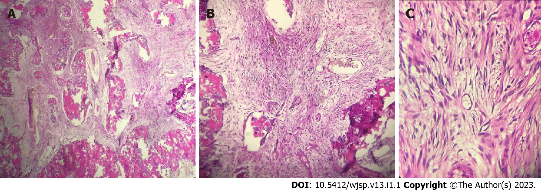
Figure 4 Pathology examination images of the excised tumor.
A and B: Tumor specimen under the microscope at × 10 magnification; C: Tumor specimen under the microscope at × 40 magnification. It showed the bone trabecular component and fibrous connective tissues with spindle-shaped cells with cigar-shaped nuclei, eosinophilic cytoplasm, and smooth chromatin.
- Citation: Christian INWS, Michael, Setiawan K. Cemento-ossifying fibroma of the left mandible: A case report. World J Surg Proced 2023; 13(1): 1-6
- URL: https://www.wjgnet.com/2219-2832/full/v13/i1/1.htm
- DOI: https://dx.doi.org/10.5412/wjsp.v13.i1.1


