Copyright
©The Author(s) 2023.
World J Surg Proced. Mar 31, 2023; 13(1): 1-6
Published online Mar 31, 2023. doi: 10.5412/wjsp.v13.i1.1
Published online Mar 31, 2023. doi: 10.5412/wjsp.v13.i1.1
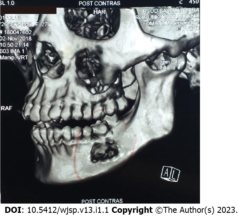
Figure 2 Computed tomography scan with 3D reconstruction revealed a circular-shaped lesion in the left mandible body with a well-defined radiolucent border, sized 2.
3 cm × 2.8 cm × 0.9 cm.
- Citation: Christian INWS, Michael, Setiawan K. Cemento-ossifying fibroma of the left mandible: A case report. World J Surg Proced 2023; 13(1): 1-6
- URL: https://www.wjgnet.com/2219-2832/full/v13/i1/1.htm
- DOI: https://dx.doi.org/10.5412/wjsp.v13.i1.1


