Copyright
©The Author(s) 2021.
World J Surg Proced. Dec 30, 2021; 11(1): 1-9
Published online Dec 30, 2021. doi: 10.5412/wjsp.v11.i1.1
Published online Dec 30, 2021. doi: 10.5412/wjsp.v11.i1.1
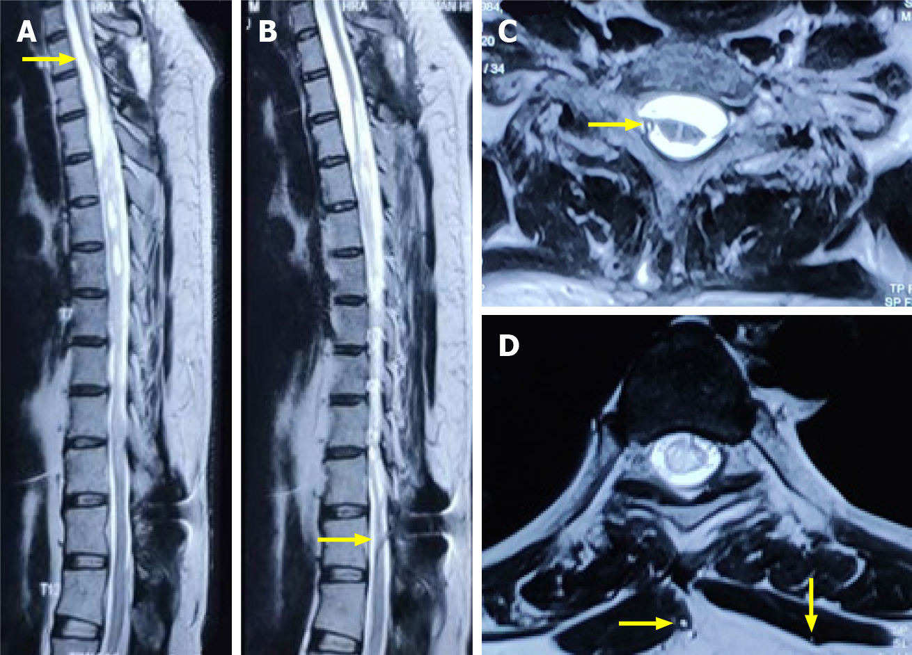
Figure 7 Magnetic resonance imaging images.
T2 weighted sagittal (A and B) and axial magnetic resonance imaging sequences (C and D) at a 4-mo interval showed the upper entry of conduit into the thecal space above and below the level of the syrinx (yellow arrows). No interval change was noted in the size of the syrinx, although a significant clinical and functional improvement was noted.
- Citation: Bhatjiwale M, Bhatjiwale M. Theco-thecal bypass technique elucidating a novel procedure and perspective on treatment of post-arachnoiditis syringomyelia: A case report. World J Surg Proced 2021; 11(1): 1-9
- URL: https://www.wjgnet.com/2219-2832/full/v11/i1/1.htm
- DOI: https://dx.doi.org/10.5412/wjsp.v11.i1.1


