Copyright
©The Author(s) 2021.
World J Surg Proced. Dec 30, 2021; 11(1): 1-9
Published online Dec 30, 2021. doi: 10.5412/wjsp.v11.i1.1
Published online Dec 30, 2021. doi: 10.5412/wjsp.v11.i1.1
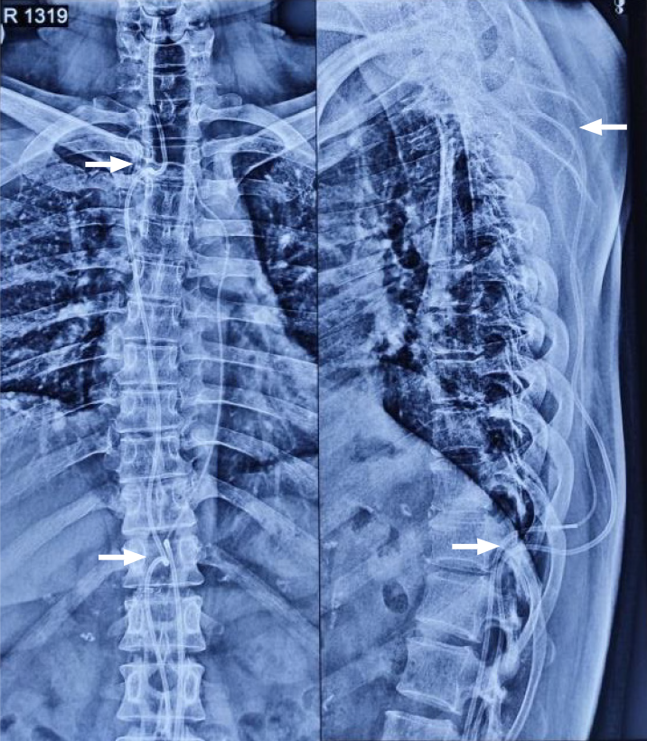
Figure 6
Postoperative plain spine radiographs (AP and lateral) confirming the intraspinal position above at D1-D2 level and below at D11 level (white arrows) along with the continuous subcutaneous track of the conduits and intrathecal portion visible.
- Citation: Bhatjiwale M, Bhatjiwale M. Theco-thecal bypass technique elucidating a novel procedure and perspective on treatment of post-arachnoiditis syringomyelia: A case report. World J Surg Proced 2021; 11(1): 1-9
- URL: https://www.wjgnet.com/2219-2832/full/v11/i1/1.htm
- DOI: https://dx.doi.org/10.5412/wjsp.v11.i1.1


