Copyright
©The Author(s) 2021.
World J Surg Proced. Dec 30, 2021; 11(1): 1-9
Published online Dec 30, 2021. doi: 10.5412/wjsp.v11.i1.1
Published online Dec 30, 2021. doi: 10.5412/wjsp.v11.i1.1
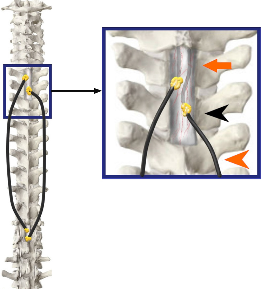
Figure 5 The tubes have been inserted into the theca on both sides of the midline approximately 7-10 mm apart (orange arrowhead).
An adequate intradural length is ensured (orange arrow) and fat grafts are lowered (black arrowhead) over a fibrin seal to augment the sealing (Inset Image).
- Citation: Bhatjiwale M, Bhatjiwale M. Theco-thecal bypass technique elucidating a novel procedure and perspective on treatment of post-arachnoiditis syringomyelia: A case report. World J Surg Proced 2021; 11(1): 1-9
- URL: https://www.wjgnet.com/2219-2832/full/v11/i1/1.htm
- DOI: https://dx.doi.org/10.5412/wjsp.v11.i1.1


