Copyright
©The Author(s) 2021.
World J Surg Proced. Dec 30, 2021; 11(1): 1-9
Published online Dec 30, 2021. doi: 10.5412/wjsp.v11.i1.1
Published online Dec 30, 2021. doi: 10.5412/wjsp.v11.i1.1
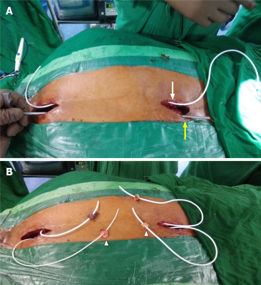
Figure 4 Connecting passages created by subcutaneous tunneling with the help of an adult ventriculoperitoneal shunt tunneller.
A: One after another, both conduits were positioned in the subcutaneous plane on either side of the midline, using an adult ventriculoperitoneal shunt tunneller. The right conduit is seen in place (white arrow) and the tunneller is in use to position the left shunt (yellow arrow); B: Both conduits were placed in the subcutaneous plane and emerged on either side of the incision with fat pads (arrowheads) placed a few centimeters from the tip to help seal the dural entry.
- Citation: Bhatjiwale M, Bhatjiwale M. Theco-thecal bypass technique elucidating a novel procedure and perspective on treatment of post-arachnoiditis syringomyelia: A case report. World J Surg Proced 2021; 11(1): 1-9
- URL: https://www.wjgnet.com/2219-2832/full/v11/i1/1.htm
- DOI: https://dx.doi.org/10.5412/wjsp.v11.i1.1


