Copyright
©The Author(s) 2017.
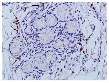
Figure 2 Macrophages in autoimmune lesions.
CD68+ macrophages infiltrate in the salivary gland tissue from SS patients. Immunohistochemical analysis using paraffin-embedded sections from lip biopsy materials was performed by staining with anti-CD68 antibody (DAKO). Biotinylated antibody and horseradish peroxidase (HRP)-conjugated streptavidin (LSAB kit, DAKO) was used as a secondary antibody, and then CD68+ cells were detected by using 3,3’-Diaminobenzidine (DAB) as a substrate. Counter staining was performed with hematoxylin. Original magnificaion is × 400. Arrows show CD68+ macrophages.
- Citation: Ushio A, Arakaki R, Yamada A, Saito M, Tsunematsu T, Kudo Y, Ishimaru N. Crucial roles of macrophages in the pathogenesis of autoimmune disease. World J Immunol 2017; 7(1): 1-8
- URL: https://www.wjgnet.com/2219-2824/full/v7/i1/1.htm
- DOI: https://dx.doi.org/10.5411/wji.v7.i1.1


