Copyright
©The Author(s) 2016.
World J Immunol. Jul 27, 2016; 6(2): 105-118
Published online Jul 27, 2016. doi: 10.5411/wji.v6.i2.105
Published online Jul 27, 2016. doi: 10.5411/wji.v6.i2.105
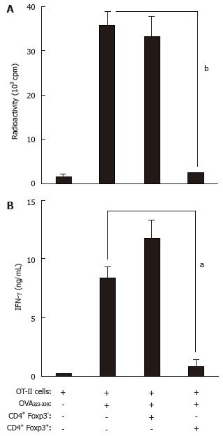
Figure 8 Treg cells from urothelium-ovalbuminGFP-Foxp3/OT-II mice are suppressive to ovalbumin-specific CD4+ T cells.
A: OT-II splenocytes were incubated alone or in the presence of OVA323-339 peptide (10 μg/mL), GFP-positive (Foxp3+) CD4+ T cells (at a 1:1 ratio), and/or GFP-negative (Foxp3-) CD4+ T cells (at a 1:1 ratio) sorted from URO-OVAGFP-Foxp3/OT-II mice for 3 d. Proliferation was assessed by labeling the cultures with 3H-thymidine for the final 18 h. Data are presented as the mean ± SD from triplicate cultures. bP < 0.001 compared with OT-II cells stimulated with OVA323-339 peptide alone (two-tailed Student’s t test); B: Culture supernatants from a parallel plate were collected after 3-d incubation and analyzed for IFN-γ by ELISA. Data are presented as the mean ± SD from duplicate cultures. aP < 0.05 compared with OT-II cells stimulated with OVA323-339 peptide alone (two-tailed Student’s t test). UROGFP-Foxp3/OT-II: Urothelium-ovalbuminGFP-Foxp3/OT-II mice.
- Citation: Liu WJ, Luo Y. Regulatory T cells suppress autoreactive CD4+ T cell response to bladder epithelial antigen. World J Immunol 2016; 6(2): 105-118
- URL: https://www.wjgnet.com/2219-2824/full/v6/i2/105.htm
- DOI: https://dx.doi.org/10.5411/wji.v6.i2.105


