Copyright
©The Author(s) 2016.
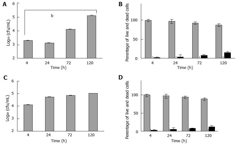
Figure 5 Mast cells infected in vitro with Mycobacterium marinum: Viable counts and cell viability.
Bone marrow derived mast cells (A, B) and human mast cell line (C, D) were infected with Mycobacterium marinum at MOI 0.5 for up to 120 h. After 4 h of infection, cells were rinsed and treated with 200 µg/mL amikacin for 2 h. At each time point intracellular bacteria were determined by enumerating colony forming units (CFU) (A, C). The values are mean ± SD of three to four independent experiments, each in triplicate. A: P < 0.0008. Viability was assessed for mast cells at these timepoints (B, D). After 4 h of infection, the cells were washed and treated with 200 μg/mL amikacin for 2 h. The values are mean ± SD of three independent experiments, each in triplicate. MOI: Multiplicity of infection.
- Citation: Siad S, Byrne S, Mukamolova G, Stover C. Intracellular localisation of Mycobacterium marinum in mast cells. World J Immunol 2016; 6(1): 83-95
- URL: https://www.wjgnet.com/2219-2824/full/v6/i1/83.htm
- DOI: https://dx.doi.org/10.5411/wji.v6.i1.83


