Copyright
©The Author(s) 2016.
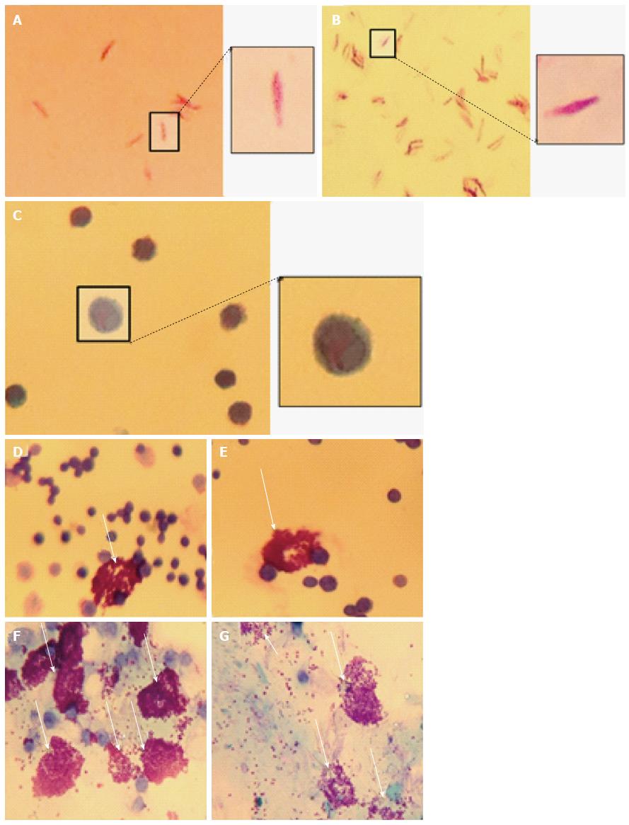
Figure 3 Peritoneal mast cells stimulated with Mycobacterium marinum degranulate.
Heat-killed (A) and untreated (B) Mycobacterium marinum (M. marinum) positive for Kinyoun’s acid fast stain, while peritoneal mast cells from mice injected with heat-killed M. marinum were negative (C). Metachromatic granules of peritoneal mast cells are stained with toluidine blue from unstimulated mice (D, E) and mice injected with heat-killed M. marinum (F, G). An exemplary image of each experimental mouse is shown (Olympus CH2, × 100 oil immersion, captured using iPhone 4S).
- Citation: Siad S, Byrne S, Mukamolova G, Stover C. Intracellular localisation of Mycobacterium marinum in mast cells. World J Immunol 2016; 6(1): 83-95
- URL: https://www.wjgnet.com/2219-2824/full/v6/i1/83.htm
- DOI: https://dx.doi.org/10.5411/wji.v6.i1.83


