Copyright
©2014 Baishideng Publishing Group Inc.
World J Immunol. Nov 27, 2014; 4(3): 174-184
Published online Nov 27, 2014. doi: 10.5411/wji.v4.i3.174
Published online Nov 27, 2014. doi: 10.5411/wji.v4.i3.174
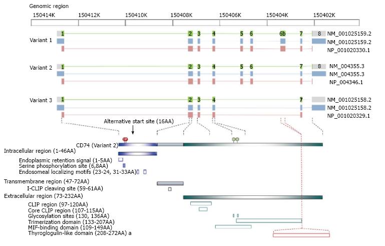
Figure 1 The variants and the corresponding protein structures of cluster of differentiation 74.
The upper panel illustrates the corresponding position among genomic region, NCBI reference sequence number and reference protein accession, exon (green box) and intron (green line) localization of DNA, transcripts (light blue box), and protein (pink box) of the three cluster of differentiation 74 (CD74) variants. The lower panel, CD74 variants contains three regions including intracellular, transmembrane and extracellular regions with the indicated functional domains and identified residues for post-translational modification. Variant 2 transcripts two isoforms, p33 and p35 caused by the alternative start site. Variant 1 transcripts two isoforms, p41 and p43, with an exon 6b-encoded thyroglobulin type I domain to interact with cathepsin. Variant 3 lacks exon 5, 6, and 6b, which translates truncated trimerization domain and truncated macrophage migration inhibitory factor (MIF)-binding domain and remains only the CLIP region to function as major histocompatibility complex class II mask. The amino acid residues refer to human variant 2 (p35).
- Citation: Liu YH, Lin JY. Recent advances of cluster of differentiation 74 in cancer. World J Immunol 2014; 4(3): 174-184
- URL: https://www.wjgnet.com/2219-2824/full/v4/i3/174.htm
- DOI: https://dx.doi.org/10.5411/wji.v4.i3.174


