Copyright
©2013 Baishideng Publishing Group Co.
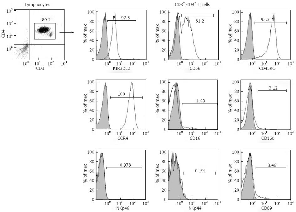
Figure 2 Immunostainings of the CD4+ T lymphocyte population.
Cells were positive for KIR3DL2, CD56, CD45RO and CCR4, but negative for the NK cell markers CD16, CD160 and NKp46 and for the activation markers NKp44 and CD69. Analyses were performed on a FC500 flow cytometer (Beckman Coulter).
- Citation: Thonnart N, Ram-Wolff C, Bagot M, Bensussan A, Marie-Cardine A. Aberrant expression of CD56 by circulating Sézary syndrome malignant T lymphocytes. World J Immunol 2013; 3(3): 68-71
- URL: https://www.wjgnet.com/2219-2824/full/v3/i3/68.htm
- DOI: https://dx.doi.org/10.5411/wji.v3.i3.68


