Copyright
©2013 Baishideng Publishing Group Co.
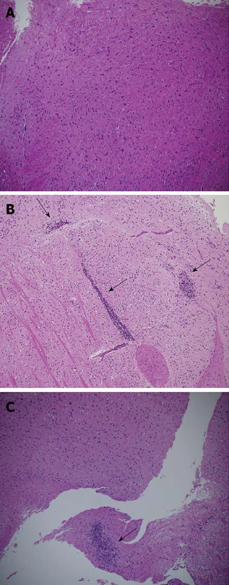
Figure 4 The extent of cellular infiltration in anti-eo-2 mAb treated experimental autoimmune encephalomyelitis mice is low compared with PBS treated experimental autoimmune encephalomyelitis mice and their healthy littermates.
A: Hematoxylin and eosin staining of representative brain sections from healthy C57BL/6 mice;B: PBS treated experimental autoimmune encephalomyelitis (EAE) mice; C: EAE mice treated with 100 μg D8. Lower extent of cellular infiltration in D8 100 μg treated group is observed compared with the IgG treated group. Arrows indicate inflammatory infiltration. Magnification × 200.
- Citation: Mausner-Fainberg K, Karni A, George J, Entin-Meer M, Afek A. Eotaxin-2 blockade ameliorates experimental autoimmune encephalomyelitis. World J Immunol 2013; 3(1): 7-14
- URL: https://www.wjgnet.com/2219-2824/full/v3/i1/7.htm
- DOI: https://dx.doi.org/10.5411/wji.v3.i1.7


