Copyright
©The Author(s) 2015.
World J Clin Urol. Nov 24, 2016; 5(3): 93-96
Published online Nov 24, 2016. doi: 10.5410/wjcu.v5.i3.93
Published online Nov 24, 2016. doi: 10.5410/wjcu.v5.i3.93
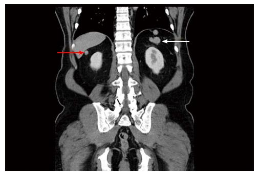
Figure 2 Coronal computed tomography image of the pelvis.
White arrow pointing to nodular soft tissue mass in left upper quadrant, where discrete spleen was not visualised; red arrow pointing to soft tissue mass beneath liver.
- Citation: Foreman D, Plagakis SA. Splenunculi mimicking metastases in a patient with locally advanced prostate cancer. World J Clin Urol 2016; 5(3): 93-96
- URL: https://www.wjgnet.com/2219-2816/full/v5/i3/93.htm
- DOI: https://dx.doi.org/10.5410/wjcu.v5.i3.93


