Copyright
©2014 Baishideng Publishing Group Inc.
World J Clin Urol. Nov 24, 2014; 3(3): 325-329
Published online Nov 24, 2014. doi: 10.5410/wjcu.v3.i3.325
Published online Nov 24, 2014. doi: 10.5410/wjcu.v3.i3.325
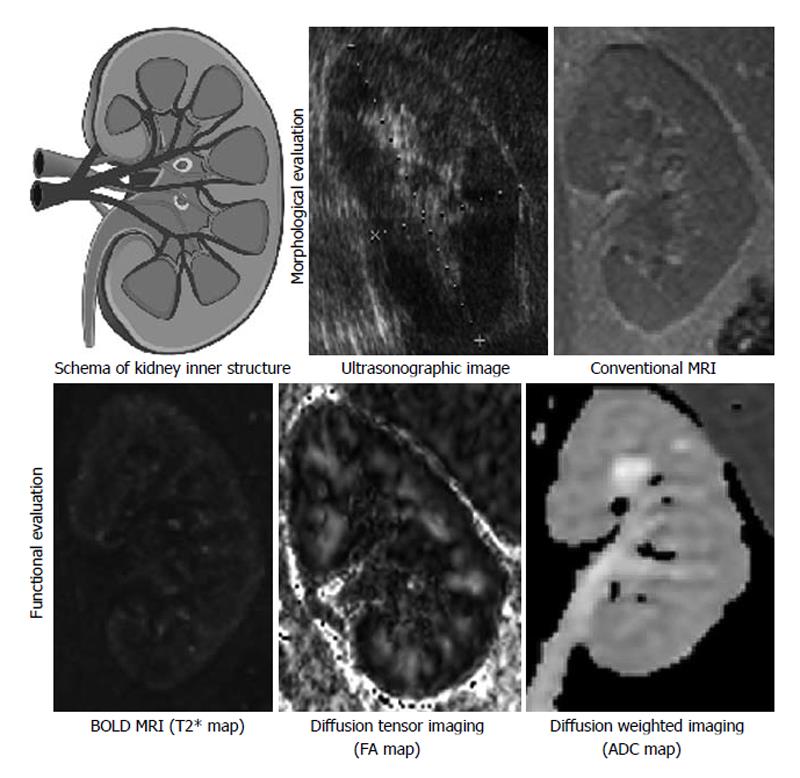
Figure 1 Various modalities for kidney.
It is noteworthy that the cortex and medulla are distinguishable on the basis of function or microstructure in functional MRI. For example, in T2* map of BOLD MRI, the medulla was seen darker than the cortex because of the difference in amount of deoxy-hemoglobin; in FA map of diffusion tensor imaging, FA of medulla was higher than the cortex probably because straight microstructure of medulla including distal tubules and surrounding blood capillaries restrict irregularity of Brownian movement of protons. Contrast and angle are adjusted respectively. Each of the kidney images is of a different patient. BOLD: Blood oxygenation level-dependent; MRI: Magnetic resonance imaging; FA: Fractional anisotropy; ADC: Apparent diffusion coefficient.
- Citation: Inoue T, Kozawa E, Okada H, Suzuki H. Morphological and functional evaluation of chronic kidney disease using magnetic resonance imaging. World J Clin Urol 2014; 3(3): 325-329
- URL: https://www.wjgnet.com/2219-2816/full/v3/i3/325.htm
- DOI: https://dx.doi.org/10.5410/wjcu.v3.i3.325


