Copyright
©2014 Baishideng Publishing Group Inc.
World J Clin Urol. Nov 24, 2014; 3(3): 310-319
Published online Nov 24, 2014. doi: 10.5410/wjcu.v3.i3.310
Published online Nov 24, 2014. doi: 10.5410/wjcu.v3.i3.310
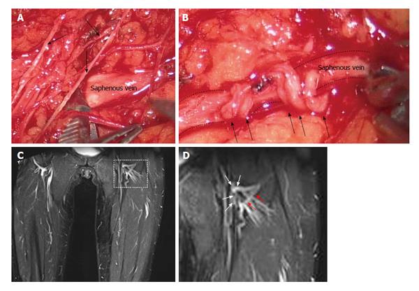
Figure 5 Lymphatic meshes were observed not only in patients who underwent spermatic funicular microsurgery, but also in patients who positively responded to the treatment of the lower limb swelling.
A: Microsurgical identification of the saphenous vein and of lymphatic vessels (arrows) in the crural area; B: Latero-terminal lympho-venous anastomosis, confectioned with interrupted stitches, between lymphatic vessels (arrows) and the saphenous vein (dashed line) in proximity of the femoral-saphenous cross; C: Magnetic resonance image of the lower limb obtained in the same patient of A, B at 8 years after microsurgery. Details of the left limb crural area (delimited by the dashed line) are presented in D; D: White arrows identify the sites were latero-terminal lympho-venous anastomosis were confectioned, while red arrows focus on a new developed lymphatic collector.
- Citation: Mukenge SM, Negrini D, Catena M, Ferla G. Innovative microsurgical treatment of male external genitals lymphedema. World J Clin Urol 2014; 3(3): 310-319
- URL: https://www.wjgnet.com/2219-2816/full/v3/i3/310.htm
- DOI: https://dx.doi.org/10.5410/wjcu.v3.i3.310


