Copyright
©The Author(s) 2017.
World J Clin Pediatr. Feb 8, 2017; 6(1): 52-59
Published online Feb 8, 2017. doi: 10.5409/wjcp.v6.i1.52
Published online Feb 8, 2017. doi: 10.5409/wjcp.v6.i1.52
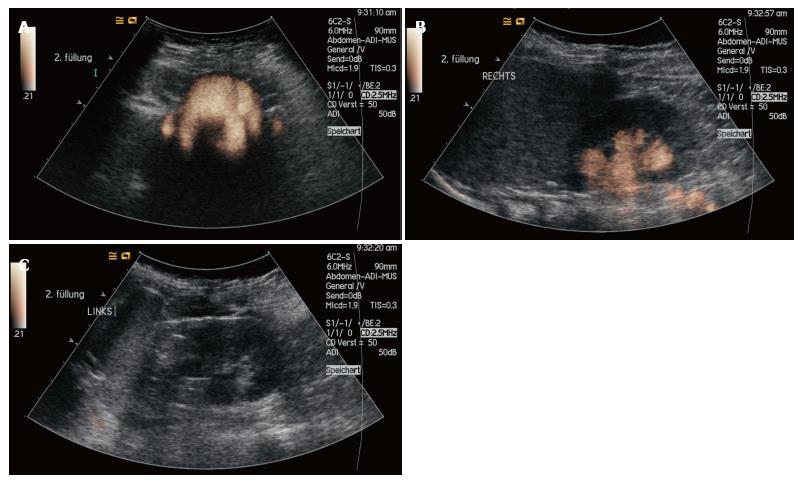
Figure 2 Voiding urosonography of a 6-year-old girl with proven, bilateral vesicoureteral reflux (grade I left and grade III right).
The voiding urosonography scan shows the urosonography contrast agent microbubbles in both ureters and in the left renal pelvis (A: Bladder and distal ureters; B: Right kidney and proximal ureter; C: Left kidney).
- Citation: Sauer A, Wirth C, Platzer I, Neubauer H, Veldhoen S, Dierks A, Kaiser R, Kunz A, Beer M, Bley T. Off-label-use of sulfur-hexafluoride in voiding urosonography for diagnosis of vesicoureteral reflux in children: A survey on adverse events. World J Clin Pediatr 2017; 6(1): 52-59
- URL: https://www.wjgnet.com/2219-2808/full/v6/i1/52.htm
- DOI: https://dx.doi.org/10.5409/wjcp.v6.i1.52


