Copyright
©The Author(s) 2017.
World J Clin Pediatr. Feb 8, 2017; 6(1): 103-109
Published online Feb 8, 2017. doi: 10.5409/wjcp.v6.i1.103
Published online Feb 8, 2017. doi: 10.5409/wjcp.v6.i1.103
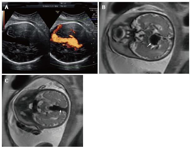
Figure 1 A 42-year-old G2P1001 presented for a routine growth ultrasound at 36 wk 5 d.
A: Prenatal ultrasonography shows vein of Galen aneurysm and color flow examination reveals a turbulence flow in the lesion and in the connected strait sinus; B: Prenatal T1-weighted magnetic resonance images show the markedly enlarged median procencephalic vein of Markowsky, characteristic of vein of Galen aneurysmal malformation (arrow); C: Persistence of the falcine draining sinus (arrow).
- Citation: Puvabanditsin S, Mehta R, Palomares K, Gengel N, Silva CFD, Roychowdhury S, Gupta G, Kashyap A, Sorrentino D. Vein of Galen malformation in a neonate: A case report and review of endovascular management. World J Clin Pediatr 2017; 6(1): 103-109
- URL: https://www.wjgnet.com/2219-2808/full/v6/i1/103.htm
- DOI: https://dx.doi.org/10.5409/wjcp.v6.i1.103


