Copyright
©The Author(s) 2017.
World J Clin Pediatr. Feb 8, 2017; 6(1): 10-23
Published online Feb 8, 2017. doi: 10.5409/wjcp.v6.i1.10
Published online Feb 8, 2017. doi: 10.5409/wjcp.v6.i1.10
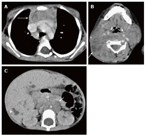
Figure 22 Thymic involvement in systemic Langerhans cell histiocytosis in a 2-year-old girl.
CECT axial section at the level of the arch of the aorta (A) shows heterogenous appearance of the thymus with a nodular outline (arrow). Axial section of the suprahyoid neck (B) shows multiple bilateral enlarged lymph nodes with cystic change (arrow). CECT of the abdomen (C) reveals hepatomegaly with multiple small focal hypodense lesions (arrowhead) in both lobes of the liver and enlarged retroperitoneal lymph nodes (arrow).
- Citation: Manchanda S, Bhalla AS, Jana M, Gupta AK. Imaging of the pediatric thymus: Clinicoradiologic approach. World J Clin Pediatr 2017; 6(1): 10-23
- URL: https://www.wjgnet.com/2219-2808/full/v6/i1/10.htm
- DOI: https://dx.doi.org/10.5409/wjcp.v6.i1.10


