Copyright
©The Author(s) 2017.
World J Clin Pediatr. Feb 8, 2017; 6(1): 10-23
Published online Feb 8, 2017. doi: 10.5409/wjcp.v6.i1.10
Published online Feb 8, 2017. doi: 10.5409/wjcp.v6.i1.10
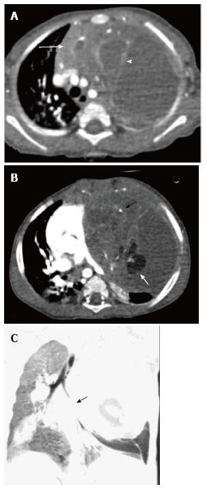
Figure 21 Immature teratoma in a 1-year-old boy.
CECT axial sections (A and B) reveal a large cystic mass in the anterior mediastinum. The mass has enhancing solid component (arrow in A), internal septations (arrowhead in A), fat (white arrow in B), and calcific foci (black arrow in B). Note the mass effect on heart and mediastinal vascular structures. Coronal minimum intensity projection image (C) shows the narrowing of left lower lobe bronchus (arrow) and basal atelectasis.
- Citation: Manchanda S, Bhalla AS, Jana M, Gupta AK. Imaging of the pediatric thymus: Clinicoradiologic approach. World J Clin Pediatr 2017; 6(1): 10-23
- URL: https://www.wjgnet.com/2219-2808/full/v6/i1/10.htm
- DOI: https://dx.doi.org/10.5409/wjcp.v6.i1.10


