Copyright
©The Author(s) 2017.
World J Clin Pediatr. Feb 8, 2017; 6(1): 10-23
Published online Feb 8, 2017. doi: 10.5409/wjcp.v6.i1.10
Published online Feb 8, 2017. doi: 10.5409/wjcp.v6.i1.10
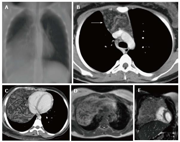
Figure 18 Thymolipoma in 17-year-old female.
Thick MPR (multiplanar reconstruction) in the coronal plane (A) shows a large anterior and middle mediastinal mass with low attenuation and sharp margins. CECT axial images (B and C) reveal a large anterior mediastinal mass lesion extending into the right hemithorax. The mass is predominantly of fat attenuation (arrow in A). Note that there is no displacement of the heart or great vessels. Axial T1WI (D) and coronal T2WI (E) show a large heterogenous fat containing mass with intervening fibrous septa. The mass has predominantly hyperintense signal on T1WI and intermediate signal on T2WI consistent with fat.
- Citation: Manchanda S, Bhalla AS, Jana M, Gupta AK. Imaging of the pediatric thymus: Clinicoradiologic approach. World J Clin Pediatr 2017; 6(1): 10-23
- URL: https://www.wjgnet.com/2219-2808/full/v6/i1/10.htm
- DOI: https://dx.doi.org/10.5409/wjcp.v6.i1.10


