Copyright
©The Author(s) 2017.
World J Clin Pediatr. Feb 8, 2017; 6(1): 10-23
Published online Feb 8, 2017. doi: 10.5409/wjcp.v6.i1.10
Published online Feb 8, 2017. doi: 10.5409/wjcp.v6.i1.10
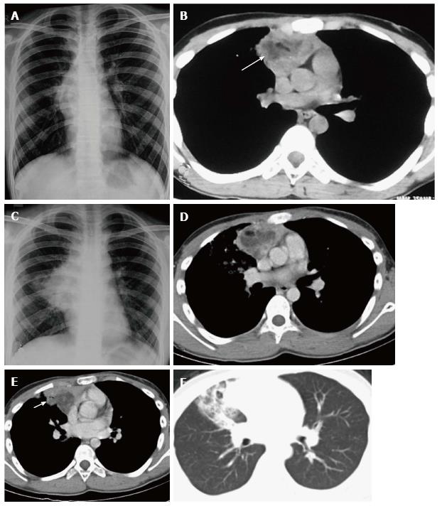
Figure 15 Ruptured anterior mediastinal teratoma in a 14-year-old male.
Chest radiograph (A) shows a lobulated soft tissue mass in the right hilar region with obscured inferior margin. CECT axial section (B) shows a heterogenous mass in the anterior mediastinum with soft tissue, fluid and fat suggestive of anterior mediastinal teratoma. The patient refused surgery and presented 5 mo later, with complaint of cough and expectoration of foul smelling material. Chest radiograph (C) shows a large right hilar mass with irregular margins and patchy consolidation in the adjacent right mid zone. CECT (D) also demonstrates the increase in the size of the mass and irregular margins. There is evidence of small air foci (arrow in E) within this mass with adjacent consolidation suggestive of rupture into the tracheobronchial tree. Axial CT lung window (F) shows the adjacent ground glass and consolidation in the right middle lobe.
- Citation: Manchanda S, Bhalla AS, Jana M, Gupta AK. Imaging of the pediatric thymus: Clinicoradiologic approach. World J Clin Pediatr 2017; 6(1): 10-23
- URL: https://www.wjgnet.com/2219-2808/full/v6/i1/10.htm
- DOI: https://dx.doi.org/10.5409/wjcp.v6.i1.10


