Copyright
©The Author(s) 2017.
World J Clin Pediatr. Feb 8, 2017; 6(1): 10-23
Published online Feb 8, 2017. doi: 10.5409/wjcp.v6.i1.10
Published online Feb 8, 2017. doi: 10.5409/wjcp.v6.i1.10
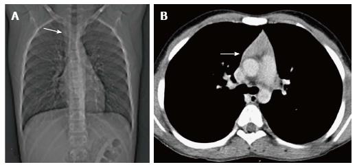
Figure 10 Thymic hyperplasia in a 12-year-old boy treated for Hodgkin’s lymphoma for 8 mo.
CT scanogram (A) shows subtle mediastinal widening (arrow). CECT axial section (B) shows mild thymic enlargement with convex margins and homogenous density (arrow) consistent with rebound hyperplasia.
- Citation: Manchanda S, Bhalla AS, Jana M, Gupta AK. Imaging of the pediatric thymus: Clinicoradiologic approach. World J Clin Pediatr 2017; 6(1): 10-23
- URL: https://www.wjgnet.com/2219-2808/full/v6/i1/10.htm
- DOI: https://dx.doi.org/10.5409/wjcp.v6.i1.10


