Copyright
©The Author(s) 2016.
World J Clin Pediatr. May 8, 2016; 5(2): 228-233
Published online May 8, 2016. doi: 10.5409/wjcp.v5.i2.228
Published online May 8, 2016. doi: 10.5409/wjcp.v5.i2.228
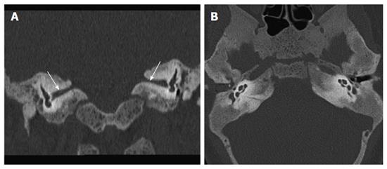
Figure 3 Coronal reformatted high resolution computed tomography.
(A) image showing thickened sclerotic bones causing narrowing of bilateral internal auditory canal (arrows); normal cochlea and vestibule seen in both ears on axial high resolution computed tomography image (B).
- Citation: Verma R, Jana M, Bhalla AS, Kumar A, Kumar R. Diagnosis of osteopetrosis in bilateral congenital aural atresia: Turning point in treatment strategy. World J Clin Pediatr 2016; 5(2): 228-233
- URL: https://www.wjgnet.com/2219-2808/full/v5/i2/228.htm
- DOI: https://dx.doi.org/10.5409/wjcp.v5.i2.228


