Copyright
©The Author(s) 2016.
World J Clin Pediatr. Feb 8, 2016; 5(1): 136-142
Published online Feb 8, 2016. doi: 10.5409/wjcp.v5.i1.136
Published online Feb 8, 2016. doi: 10.5409/wjcp.v5.i1.136
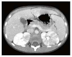
Figure 5 Case 4: Abdominal computed tomography.
Computed tomography scan of the abdomen after intravenous contrast injection reveals multiple wedge-shaped cortical hypodense lesions in the both kidneys, more represented in the left one.
- Citation: Bibalo C, Apicella A, Guastalla V, Marzuillo P, Zennaro F, Tringali C, Taddio A, Germani C, Barbi E. Acute lobar nephritis in children: Not so easy to recognize and manage. World J Clin Pediatr 2016; 5(1): 136-142
- URL: https://www.wjgnet.com/2219-2808/full/v5/i1/136.htm
- DOI: https://dx.doi.org/10.5409/wjcp.v5.i1.136


