Copyright
©The Author(s) 2016.
World J Clin Pediatr. Feb 8, 2016; 5(1): 1-15
Published online Feb 8, 2016. doi: 10.5409/wjcp.v5.i1.1
Published online Feb 8, 2016. doi: 10.5409/wjcp.v5.i1.1
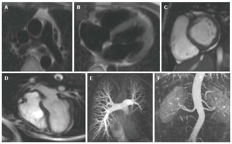
Figure 1 Examples of images produced by individual cardiovascular magnetic resonance pulse sequences.
A and B are black blood spin echo images showing the ascending and descending aorta at the main pulmonary artery level A and the 4 cardiac chambers B; C and D are bright blood SSFP cine images showing short axis and 4 chamber views respectively, while E and F are examples of 3D contrast-enhanced MRA; E is a contrast-enhanced MRA of the pulmonary tree in a patient with a large sarcoma. There is no opacification of the arterial supply of the left lower lobe, indicating complete occlusion of the lower branch of the left pulmonary artery; F is a contrast-enhanced MRA of the descending aorta for assessment of renal anatomy, demonstrating an accessory renal artery to left kidney (a common normal variant). SSFP: Steady state free precession; MRA: Magnetic resonance angiography.
- Citation: Mitchell FM, Prasad SK, Greil GF, Drivas P, Vassiliou VS, Raphael CE. Cardiovascular magnetic resonance: Diagnostic utility and specific considerations in the pediatric population. World J Clin Pediatr 2016; 5(1): 1-15
- URL: https://www.wjgnet.com/2219-2808/full/v5/i1/1.htm
- DOI: https://dx.doi.org/10.5409/wjcp.v5.i1.1


