Copyright
©The Author(s) 2015.
World J Clin Pediatr. Nov 8, 2015; 4(4): 94-105
Published online Nov 8, 2015. doi: 10.5409/wjcp.v4.i4.94
Published online Nov 8, 2015. doi: 10.5409/wjcp.v4.i4.94
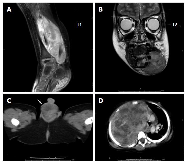
Figure 1 Radiographic images of common rhabdomyosarcoma.
A: Extremity; B: Head and neck; C: Genitourinary (paratesticular); D: Axial (intraabdominal).
- Citation: Sangkhathat S. Current management of pediatric soft tissue sarcomas. World J Clin Pediatr 2015; 4(4): 94-105
- URL: https://www.wjgnet.com/2219-2808/full/v4/i4/94.htm
- DOI: https://dx.doi.org/10.5409/wjcp.v4.i4.94


