Copyright
©The Author(s) 2015.
World J Clin Pediatr. Nov 8, 2015; 4(4): 160-166
Published online Nov 8, 2015. doi: 10.5409/wjcp.v4.i4.160
Published online Nov 8, 2015. doi: 10.5409/wjcp.v4.i4.160
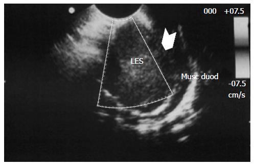
Figure 2 Endoscopic ultrasound showed an homogeneous, hypoechoic mass of 36 mm located into the echo-layer 3 (submucosa), but not completely separated from the muscularis propria (echo-layer 4); in some endoscopic ultrasound scans, the lesion showed features suggesting origin from the muscular layer, thus mimicking gastrointestinal stromal tumors features.
The wall of the lesion was homogeneous and the external part showed hyperechoic spots, suggestive of microcalcifications.
- Citation: Righetti L, Parolini F, Cengia P, Boroni G, Cheli M, Sonzogni A, Alberti D. Inflammatory fibroid polyps in children: A new case report and a systematic review of the pediatric literature. World J Clin Pediatr 2015; 4(4): 160-166
- URL: https://www.wjgnet.com/2219-2808/full/v4/i4/160.htm
- DOI: https://dx.doi.org/10.5409/wjcp.v4.i4.160


