Copyright
©The Author(s) 2015.
World J Clin Pediatr. May 8, 2015; 4(2): 30-34
Published online May 8, 2015. doi: 10.5409/wjcp.v4.i2.30
Published online May 8, 2015. doi: 10.5409/wjcp.v4.i2.30
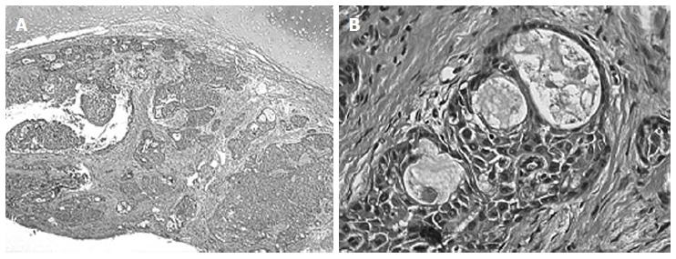
Figure 2 Case 3, mucoepidermoid carcinoma, histological findings.
A: The tumor was pathologically diagnosed as mucoepidermoid Carcinoma (HE stain x 5). Neoplastic infiltration of the bronchial submucosa characterized by a mixture of glands, tubules, cysts and solid areas; B: Glandular component of the tumor, lined by columnar mucinous cells and containing mucin, is intimately admixed with solid islands composed of squamous and intermediate cells (HE stain x 63).
- Citation: Madafferi S, Catania VD, Accinni A, Boldrini R, Inserra A. Endobronchial tumor in children: Unusual finding in recurrent pneumonia, report of three cases. World J Clin Pediatr 2015; 4(2): 30-34
- URL: https://www.wjgnet.com/2219-2808/full/v4/i2/30.htm
- DOI: https://dx.doi.org/10.5409/wjcp.v4.i2.30


