Copyright
©The Author(s) 2015.
World J Clin Pediatr. May 8, 2015; 4(2): 30-34
Published online May 8, 2015. doi: 10.5409/wjcp.v4.i2.30
Published online May 8, 2015. doi: 10.5409/wjcp.v4.i2.30
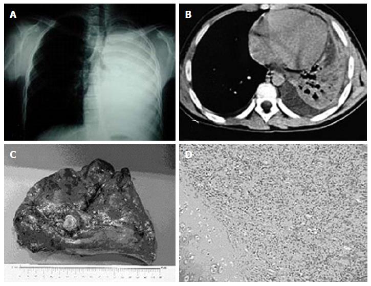
Figure 1 Case 1, endobronchial carcinoid.
A: Antero-posterior chest X-ray diagnosed at the onset of the last episode of pneumonia, demonstrating a complete collapse of the left lung with hyperdistension of controlateral one. There is also an aerial bronchogram with a block before the carena; B: Axial computed tomography-scan (CT-scan), performed before operation, demonstrating atelectasis of left lung with pleural effusion; in particular we can observe the obstruction of the left main bronchus; C: Gross findings of the resected left lung, on lateral surface a 1.5 cm polypoid mass obstructing the left main bronchus, firmly adherent to the wall and with a differing consistency from hard to elastic; D: The tumor was pathologically diagnosed as endobronchial carcinoid: on the left the cartilaginous wall and on the right the tumor (HE stain x 20). Monomorphic proliferation with cells forming pseudoacinouses patterns. Absence of atypical cells or abundant mitosis.
- Citation: Madafferi S, Catania VD, Accinni A, Boldrini R, Inserra A. Endobronchial tumor in children: Unusual finding in recurrent pneumonia, report of three cases. World J Clin Pediatr 2015; 4(2): 30-34
- URL: https://www.wjgnet.com/2219-2808/full/v4/i2/30.htm
- DOI: https://dx.doi.org/10.5409/wjcp.v4.i2.30


