Copyright
©2013 Baishideng Publishing Group Co.
World J Clin Pediatr. Nov 8, 2013; 2(4): 54-64
Published online Nov 8, 2013. doi: 10.5409/wjcp.v2.i4.54
Published online Nov 8, 2013. doi: 10.5409/wjcp.v2.i4.54
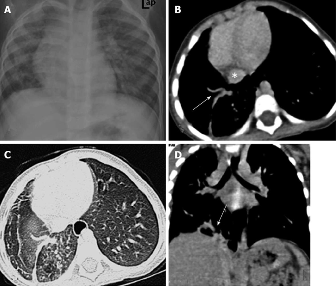
Figure 8 Pulmonary venolobar hypoplasia.
Chest radiograph (A) shows volume loss of the right hemithorax with ipsilateral mediastinal shift. Contrast-enhanced computed tomography images (B-D) showing anomalous right inferior pulmonary vein (arrows) coursing inferiorly towards the inferior vena cava (asterisk).
- Citation: Singh D, Bhalla AS, Veedu PT, Arora A. Imaging evaluation of hemoptysis in children. World J Clin Pediatr 2013; 2(4): 54-64
- URL: https://www.wjgnet.com/2219-2808/full/v2/i4/54.htm
- DOI: https://dx.doi.org/10.5409/wjcp.v2.i4.54


