Copyright
©2013 Baishideng Publishing Group Co.
World J Clin Pediatr. Nov 8, 2013; 2(4): 54-64
Published online Nov 8, 2013. doi: 10.5409/wjcp.v2.i4.54
Published online Nov 8, 2013. doi: 10.5409/wjcp.v2.i4.54
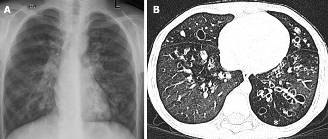
Figure 6 A 10-year-old boy with post-infectious bronchiectasis.
Frontal chest radiograph (A) showing multiple cystic lucencies and tubular opacities in both lungs. Chest high resolution CT image (B) shows multiple areas of cystic bronchiectasis with associated air trapping (asterisk).
- Citation: Singh D, Bhalla AS, Veedu PT, Arora A. Imaging evaluation of hemoptysis in children. World J Clin Pediatr 2013; 2(4): 54-64
- URL: https://www.wjgnet.com/2219-2808/full/v2/i4/54.htm
- DOI: https://dx.doi.org/10.5409/wjcp.v2.i4.54


